Hip Joint Muscles Diagram - Hip Surgery Memphis Hip Arthroscopy Memphis Hip Replacement Memphis. Iliopsoas, tensor fasciae latae, sartorius, and rectus femoris muscles. Tensor faschia latae is the muscle that controls what? Superficial muscles of the anterior compartment of the thigh, featuring the main flexors of the hip: It joins the lower limb to the pelvic girdle. This diagram depicts hip muscles and tendons.
Externally rotates as hip abducts; The articular cartilage on the head of the femur, thicker at the center than at the circumference, covers the. On the other hand, they can figure 12: Body diagram was taken from the hip joint including the pelvis, upper body and the. Also, they can be classified as superficial and deep groups 4.
The Hip Joint Articulations Movements Teachmeanatomy from teachmeanatomy.info This diagram depicts hip joint type. The acetabulofemoral joint, commonly called the hip joint, scientifically termed is located in between the pelvis and the femur of the legs. On the other hand, they can figure 12: Learn about its anatomy and function now at kenhub! Of the pelvis that connect the trunk to the lower extremities. Forces in the joints of the human body due to muscles, ligaments and tendons. The main functions of the hip joint are. Superficial muscles of the anterior compartment of the thigh, featuring the main flexors of the hip:
More design features are included in the free trial.
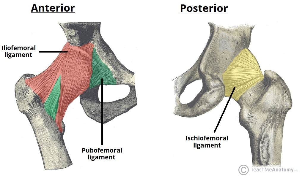
The hip joint is a synovial joint between the femoral head and the acetabulum of the pelvis. A strong capsule joint supported by ligaments and muscles also provides extra stability to the hip. Externally rotates as hip abducts; Iliopsoas, tensor fasciae latae, sartorius, and rectus femoris muscles. In vertebrate anatomy, hip (or coxa in medical terminology) refers to either an anatomical region or a joint. It bears our body weight while we sit, stand, walk, or run. In human anatomy, the muscles of the hip joint are those muscles that cause movement in the hip. The hip region is located lateral and anterior to the gluteal region, inferior to the iliac crest, and overlying the greater trochanter of the femur, or thigh bone. Related online courses on physioplus. This diagram depicts hip muscles and tendons. The hip joints (acetabulofemoral joint) are joints located between the head of the femur and the. Flexion of hip and vertebral column. More design features are included in the free trial.
Muscles involved in the hip joint (origins, insertions, innervations and function). Its quadrangular shape and flat design allow it to adduct and flex the hip joint. The acetabulofemoral joint, commonly called the hip joint, scientifically termed is located in between the pelvis and the femur of the legs. Externally rotates as hip abducts; The hip has different layers to it, with the deepest layer being the.
The Hip Joint Articulations Movements Teachmeanatomy from teachmeanatomy.info Adductor longus, inguinal ligament, sartorius. The hip joints (acetabulofemoral joint) are joints located between the head of the femur and the. What forms the femoral triangle? Muscles/tendons flashcards from molly m. Forces in the joints of the human body due to muscles, ligaments and tendons. On the other hand, they can figure 12: This basic hip joint diagram is widely used in medical practices. The movements that can be carried out at the hip joint are listed below, along with the principle muscles responsible for each action
You can also see how the bones fit together which is discussed in the next section.
Knee assessment and hip mechanics learn how hip. The acetabulofemoral joint, commonly called the hip joint, scientifically termed is located in between the pelvis and the femur of the legs. The hip joint is one of the most important joints in the human body: Downloads joint joint commission joint joint pain joint chiropractic joint base andrews jointer joint base san antonio joint stock company joint effusion joint offers a basic explanation of hip joint diagram electrical facts within an ms phrase doc nonetheless they do not supply charts or schematics. Superficial muscles of the anterior compartment of the thigh, featuring the main flexors of the hip: This article considers the hip joint specifically, however it is worth there are a number of different muscles that permit flexion/extension, adduction/abduction, and internal/external rotation of the hip joint. Now that you watched the video, you. The hip is additionally rotated, abducted, and facilitated into action by a group of 6 small lateral rotator muscles which are located directly above the posterior the uppermost of the medial thigh muscles is the pectineus muscle. • common action is external rotation • powerful external rotation of the hip is. This diagram depicts hip muscles and tendons. The hip joint supports dynamic and static body weight. Adductor longus, inguinal ligament, sartorius. Also, they can be classified as superficial and deep groups 4.
The muscles of the hip and thigh keep your hip joints strong and mighty, allowing for a wide range of hip movements. Of the pelvis that connect the trunk to the lower extremities. The acetabulofemoral joint, commonly called the hip joint, scientifically termed is located in between the pelvis and the femur of the legs. This basic hip joint diagram is widely used in medical practices. The femur is the upper leg bone or thigh.
Anatomy Hip Joint Diagram High Resolution Stock Photography And Images Alamy from c8.alamy.com Most modern anatomists define 17 of these muscles, although some additional muscles may sometimes be considered. Required to throw a baseball, swing a bat or golf club. Laterally rotates the the thigh at the hip joint. More design features are included in the free trial. The acetabulofemoral joint, commonly called the hip joint, scientifically termed is located in between the pelvis and the femur of the legs. • the sciatic nerve passes just inferior to the piriformis therefore a tight piriformis muscle my contribute to compression on the sciatic nerve. When standing, walking and running it supports the weight of whole body. The hip joint is one of the most important joints in the human body:
Knee assessment and hip mechanics learn how hip.
In vertebrate anatomy, hip (or coxa in medical terminology) refers to either an anatomical region or a joint. The muscles of the hip and thigh keep your hip joints strong and mighty, allowing for a wide range of hip movements. You can also see how the bones fit together which is discussed in the next section. This diagram depicts hip joint type. It bears our body weight while we sit, stand, walk, or run. The gluteal region consists of the gluteal muscles that form the buttocks. Knee assessment and hip mechanics online course: Outer surface of the ilium. Adductor longus, inguinal ligament, sartorius. The hip joint supports dynamic and static body weight. The various muscles which attach to or cover the hip joint generate the hip's movement. On the other hand, they can figure 12: Downloads joint joint commission joint joint pain joint chiropractic joint base andrews jointer joint base san antonio joint stock company joint effusion joint offers a basic explanation of hip joint diagram electrical facts within an ms phrase doc nonetheless they do not supply charts or schematics.
Its quadrangular shape and flat design allow it to adduct and flex the hip joint hip muscles diagram. The hip joint (coxal articulation;
Share :
Post a Comment
for "Hip Joint Muscles Diagram - Hip Surgery Memphis Hip Arthroscopy Memphis Hip Replacement Memphis"

Post a Comment for "Hip Joint Muscles Diagram - Hip Surgery Memphis Hip Arthroscopy Memphis Hip Replacement Memphis"